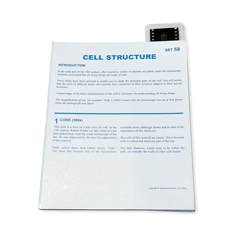
Supertek Microslide, Cell Structure, 35mm
$11.99
In stock
• Simply insert the slide and focus with the Microslide Viewer (sold separately) which is designed for use with Microslides
• Perfect for classrooms or home studies!
• Features 8 (35mm) slides of magnified specimens
Description
This Microslide, Cell Structure, includes a slide featuring 8 related 35mm images photographed through a microscope called photomicrographs. Arrows and call outs help the student locate important features. This Microslide is accompanied by a detailed lesson plan designed to stimulate, inform and question students about the topic under study, and has a pocket in which the Microslide is stored. The following magnified specimens are depicted on the slide: Cork (wm, 300X), Onion skin (wm, st, 200X), Green leaf (cs, st, 300X), Cheek cells (wm, st, 900X), Blood cells (sm, st, 900X), Nerve cells (cs, st, 300X), Bacteria (sm, st, 1,500X), and Virus (wm, 50,000X).
Additional information
| Weight | 0.07 lbs |
|---|---|
| Dimensions | 8.38 × 6.5 in |
You must be logged in to post a review.
Specifications
Unit of Measure: EachCountry of Origin: IND
Color: Black, Blue, White
Material: Paper, Film
Assembly Required: N
Package Contains: 1 Slide, 1 Lesson Plan
Package Count: 2
Battery Included: N
Barcode: 656727862232
HTS USCode: 9023000000


Reviews
There are no reviews yet.